
Japanese fMRI Research
Foot Reflexology and Japanese fMRI Research
by Barbara & Kevin Kunz
Basic precepts of reflexology have once again been upheld by a functional Magnetic Resonance Imaging (fMRI) study. Sensory stimulation applied to specific reflex areas activated specific parts of the brain correlated with the reflex area. The eye, shoulder and small intestine reflex areas of the left foot were recently tested by Japanese researchers and reaffirmed previous findings in three studies by researchers from Hong Kong. The findings of the reflexology’s theory and intended use.
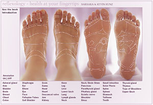
In the recent Japanese study, pressure was applied by a wooden stick to eye, shoulder and small intestine reflex areas of the left foot. Each showed activation of parts of the brain for the left foot on both the left and right sides of the brain.
Additional areas of the brain were activated and related to the part of the body reflected by the reflex area:
- Sensory stimulation applied to the eye reflex area activated the part of the brain for “processing of tactile sensations of the eye or neighboring area.”
- X
- “… our results indicate that sensory stimulation of the reflex area corresponding to the SI (small intestine) was related to a tactile sensation of the trunk. In addition its perception was not neces- sarily limited to visceral sensation and may include cutaneous sensation in the trunk.”
- X
- Stimulation applied to the shoulder reflex area showed “no specific activation, only a tendency for activation in the right (side of the brain)… location of which was most consistent with the projection of the tactile sensation to the upper limbs.
“A statistical analysis showed that reflexological stimulation of the foot reflex areas corresponding to the eye, shoulder, and small intestine activated not only the somatosensory areas corresponding to the foot, but also the somatosensory areas corresponding to the eye, shoulder, and small intestine or neighboring body parts. Thus, the findings showed that reflexological stimulation induced a somatosensory process corresponding to the stimulated reflex area and that a neuroimaging approach can be used to examine the basis of reflexology effects.” Nakamaru T, Miura N, Fukushima A, Kawashima R. “Somatotopical relationships between corti- cal activity and reflex areas in reflexology: A functional magnetic resonance imaging study,” Neuroscience Letter 2008 Oct 14 Tohoku University School of Medicine, Sendai, Japan; Department of Functional Brain Imaging, Institute of Development, Aging and Cancer (IDAC), Tohoku University, Sendai, Japan. PMID (PubMedIdentification): 18938220
(Note: Somatosensory is defined as “sensory signals from all tissues of the body including skin, viscera, muscles and joints” (The Ohio State University Medical Center))